Interactive Corporate Presentation
This document is a corporate presentation for Kodiak, an ophthalmology medicines company, dated September 2026.
THE OPHTHALMOLOGY MEDICINES COMPANY Corporate Presentation September 2026
2 Forward-Looking Statements This presentation may contain forward-looking statements within the meaning of the Private Securities Litigation Reform Act of 1995 and other federal securities laws, including statements regarding Kodiak’s plans, commitments, aspirations and goals related to Kodiak’s drug candidates. Actual results may differ materially from those indicated in the forward-looking statements as a result of various important factors which are discussed in the section entitled “Risk Factors” in Kodiak’s most recent periodic report filed with the U.S. Securities and Exchange Commission (“SEC”) as well as discussions of potential risks, uncertainties, and other important factors in Kodiak’s subsequent filings with the SEC. All information in this presentation is as of the date presented, and Kodiak undertakes no duty to update such information unless required by law. Kodiak ® , Kodiak Sciences ® , ABC ® , ABC Platform , VETi and the Kodiak logo are registered trademarks or trademarks of Kodiak Sciences Inc. in various global jurisdictions.
3 Kodiak is emerging into a fully integrated Vision Sciences company VETi: Visual Engagement Technology and imager
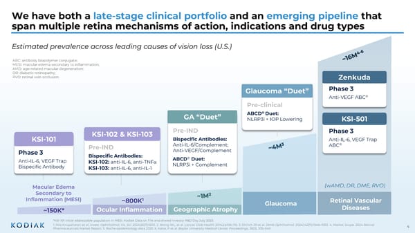
4 We have both a late-stage clinical portfolio and an emerging pipeline that span multiple retina mechanisms of action, indications and drug types ABC: antibody biopolymer conjugate; MESI: macular edema secondary to inflammation; AMD: age-related macular degeneration; DR: diabetic retinopathy; RVO: retinal vein occlusion Retinal Vascular Diseases Glaucoma Geographic Atrophy Ocular Inflammation Macular Edema Secondary to Inflammation (MESI) ~800K 1 ~150K* Phase 3 Anti-IL-6, VEGF Trap ABC ® KSI-501 Pre-clinical ABCD ® Duet: NLRP3i + IOP Lowering Glaucoma “Duet” Zenkuda Phase 3 Anti-VEGF ABC ® GA “Duet” Pre-IND Bispecific Antibodies: KSI-102: anti-IL-6, anti-TNF α KSI-103: anti-IL-6, anti-IL-1 KSI-102 & KSI-103 Estimated prevalence across leading causes of vision loss (U.S.) (wAMD, DR, DME, RVO) 1. Nila Kirupaharan et al. Invest. Ophthalmol. Vis. Sci. 2024;65(7):6510. 2. Wong WL, et al. Lancet Glob Health 2014;2:e106–116. 3. Ehrlich JR et al. JAMA Ophthalmol. 2024;142(11):1046–1053. 4. Market Scope. 2024 Retinal Pharmaceuticals Market Report. 5. Roche epidemiology data 2025. 6. Kalva, P et al. Baylor University Medical Center Proceedings , 36 (3), 335–340 *KSI-101 initial addressable population in MESI. Kodiak Data on File and shared Investor R&D Day July 2025. KSI-101 Phase 3 Anti-IL-6, VEGF Trap Bispecific Antibody ABCD ® Duet: NLRP3i + Complement Pre-IND Bispecific Antibodies: Anti-IL-6/Complement; Anti-VEGF/Complement
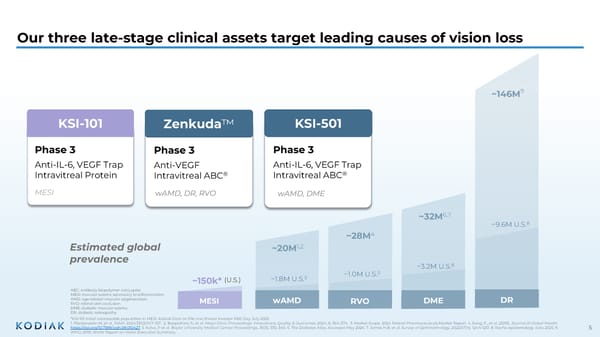
5 Our three late-stage clinical assets target leading causes of vision loss *KSI-101 initial addressable population in MESI. Kodiak Data on File and shared Investor R&D Day July 2025. 1. Fleckenstein M, et al. JAMA. 2024;331(2):147–157. 2. Boopathiraj N, et al. Mayo Clinic Proceedings: Innovations, Quality & Outcomes , 2024; 8, 364-374. 3. Market Scope. 2024 Retinal Pharmaceuticals Market Report. 4. Song, P., et al. (2019). Journal of Global Health . https://doi.org/10.7189/jogh.09.010427. 5. Kalva, P et al. Baylor University Medical Center Proceedings , 36 (3), 335–340. 6. The Diabetes Atlas, Accessed May 2026. 7. James H.B. et al. Survey of Ophthalmology, 2022;67(4): 1244-1251. 8. Roche epidemiology data 2025. 9. WHO, 2019, World Report on Vision Executive Summary. ABC: antibody biopolymer conjugate; MESI: macular edema secondary to inflammation; AMD: age-related macular degeneration; RVO: retinal vein occlusion DME: diabetic macular edema DR: diabetic retinopathy Estimated global prevalence DR DME RVO wAMD ~32M 6,7 ~28M 4 ~20M 1,2 MESI ~150k* (U.S.) ~1.8M U.S. 3 ~1.0M U.S. 5 ~3.2M U.S. 8 ~9.6M U.S. 8 ~146M 9 KSI-101 Phase 3 Anti-IL-6, VEGF Trap Intravitreal Protein MESI Zenkuda Phase 3 Anti-VEGF Intravitreal ABC ® wAMD, DR, RVO Phase 3 Anti-IL-6, VEGF Trap Intravitreal ABC ® KSI-501 wAMD, DME
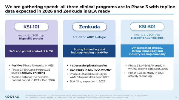
6 We are gathering speed: all three clinical programs are in Phase 3 with topline data expected in 2026 and Zenkuda is BLA ready *Based on the positive Phase 3 GLOW1 and GLOW2 studies in diabetic retinopathy, the BEACON study in retinal vein occlusion and the DAYLIGHT study in wet AMD ABC: antibody biopolymer conjugate; DR: diabetic retinopathy; RVO: retinal vein occlusion; wAMD: wet age-related macular degeneration KSI-101 Zenkuda Anti-VEGF ABC ® biologic KSI-501 Anti-IL-6, VEGF trap bispecific ABC ® biologic • Positive Phase 1b results in MESI • Phase 3 PEAK and PINNACLE studies actively enrolling • Topline data for the first 300- patient cohort in PEAK Dec. 2026 • 4 successful pivotal studies • BLA-ready in DR, RVO, wAMD* • Phase 3 DAYBREAK study in wAMD topline data Sept. 2026 • BLA filing expected in 2026 • Phase 3 DAYBREAK study in wAMD topline data Sept. 2026 • Phase 3 ALTO study in DME actively recruiting Anti-IL-6, VEGF trap bispecific protein Safe and potent control of MESI Strong immediacy and industry-leading durability Differentiated efficacy, strong immediacy and industry-leading durability
7 ZENKUDA 3Q ‘26 4Q ‘26 1Q ‘27 2Q ‘27 3Q ‘27 4Q ‘27 1Q ‘28 2Q ‘28 3Q ‘28 RVO (BEACON) DR (GLOW1) DR (GLOW2) wAMD (DAYLIGHT) wAMD (DAYBREAK) KSI-101 MESI (PEAK) MESI (PINNACLE) KSI-501 wAMD (DAYBREAK) DME (ALTO) Four Phase 3 readouts in 2026, if successful, position Kodiak to file 3 licensing applications (BLA) in 2026 and 2027 RVO: retinal vein occlusion; DR: diabetic retinopathy; AMD: age-related macular edema; DME: diabetic macular edema; MESI: macular edema secondary to inflammation; BLA: biologics license application; sBLA: supplemental biologics license application; DAYBREAK: NCT06556368; PEAK: NCT06990399; PINNACLE: NCT06996080 BLA* BLA* BLA* sBLA* • DAYBREAK topline data Sept. 2026 • Phase 3 ALTO study in DME First Patient In 3Q 2026 • Pivotal analysis 1 ( PEAK ) expected December 2026; potential BLA filing in 1H 2027 • Pivotal analysis 2 ( PEAK+PINNACLE ) expected 2Q 2027 First Patient In • DAYBREAK topline data Sept 2026; BLA filing expected in 4Q 2026 Upcoming catalysts 3 additional topline data readouts expected in 2026 4 successful pivotal studies completed 3 Licensing applications (BLA) expected in 2026 and 2027 *Timing reflects current expectations; BLA timing pending discussions with regulatory bodies, subject to change
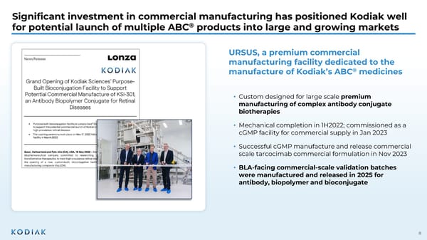
8 Significant investment in commercial manufacturing has positioned Kodiak well for potential launch of multiple ABC ® products into large and growing markets URSUS, a premium commercial manufacturing facility dedicated to the manufacture of Kodiak’s ABC ® medicines • Custom designed for large scale premium manufacturing of complex antibody conjugate biotherapies • Mechanical completion in 1H2022; commissioned as a cGMP facility for commercial supply in Jan 2023 • Successful cGMP manufacture and release commercial scale tarcocimab commercial formulation in Nov 2023 • BLA-facing commercial-scale validation batches were manufactured and released in 2025 for antibody, biopolymer and bioconjugate
Kodiak’s ABC ® Platform Science for Retina 10 ZENKUDA + KSI-501
The ABC Platform supports Kodiak’s science of immediacy and durability Designed-in Extended Tissue Residence Time A proprietary phosphorylcholine -based polymer is conjugated to an antibody to increase molecular size which extends ocular half-life High In-Vitro Potency Extended Ocular Half- Life in Animals Extended Ocular Half- Life in Humans Both unconjugated protein and conjugated protein demonstrate high binding affinity and potency in vitro 3x the ocular t½ of approved intravitreal biologics when measured in rabbits following an intravitreal injection 3x the ocular t½ of faricimab when measured from aqueous humor in patients following an intravitreal injection Powerful Immediacy via Unconjugated Antibody The unconjugated protein delivers a strong “pulse” of anti-VEGF inhibition during the loading phase, or to recapture control of the disease in patients whose disease has reactivated Extended Clinical Durability via Conjugated Antibody The conjugated protein maintains the signature durability as seen in Kodiak’s pivotal studies to date 11
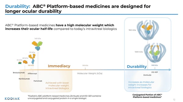
Durability: ABC ® Platform-based medicines are designed for longer ocular durability Increases as molecular weight increases for intravitreal biologics Achieved with lower molecular-weight intravitreal biologics Conjugated Portion of ABC ® Platform-based medicines* *Kodiak’s ABC platform-based medicines Zenkuda and KSI-501 combine unconjugated and conjugated protein in a single biologic Zenkuda 1000 kDa ABC ® Platform-based medicines have a high molecular weight which increases their ocular half-life compared to today’s intravitreal biologics 950 kDa KSI-501 Immediacy Durability 12
Durability: Zenkuda has a mean ocular half-life in humans of 20 days, which is 3-fold longer than faricimab 10-90th percentiles 8.3 (10th percentile) 19.8 days (Mean) 5.2 (10 th percentile) t½ <5 days t½ >11 days Faricimab Zenkuda Percent of Patients Each dot represents the ocular half-life from one individual patient. Blue dots are Zenkuda from the Phase 1b study of Zenkuda in patients with wAMD, DME and RVO. Gray dots are faricimab from Genentech, Inc. PK and ER of faricimab, Report # 1105763 0% 81% 10% 10% 0 20 40 60 80 100 Faricimab Zenkuda 31.8 (90th percentile) 11.2 (90 th percentile) 7.5 days (Mean) 10-90th percentiles Human Ocular Half-Life (Days) ~3x longer than faricimab 13
Durability: Zenkuda’s ocular half-life in humans was calculated by measuring aqueous humor concentrations over time from patients in the Phase 1b Study 1 10 100 1000 10000 100000 0 4 8 12 16 20 24 28 • Aqueous humor samples were collected from 47 subjects in the Zenkuda Phase 1b study in patients with wet AMD, DME and RVO and were used to evaluate Zenkuda ocular half life in patients • Aqueous humor samples were collected at baseline and at Week 4, 12, 14, 16, 18, 20 and 24 and measured for Zenkuda concentrations • Samples collected between the last loading dose and the next re-dose were used to determine ocular half-life of Zenkuda Using Aqueous Humor Concentration of Zenkuda to Determine Ocular t 1/2 in Human Weeks Zenkuda aqueous humor concentration (ng/ml) 3 loading doses of intravitreal injections of 5 mg Zenkuda AH samples from Week 12 – 24 were used to determine Zenkuda t 1/2 Data from one individual patient from the Phase 1b study of Zenkuda is used to illustrate the methodology here. Patients received as-needed redosing after week 8 and aqueous humor samples collected after a re-dose were excluded from the analysis 14
12 16 20 24 1 10 100 1000 10000 100000 Durability: Zenkuda achieved an extended ocular half-life of >20 days in 45% of sampled patients from the Phase 1b Study All Patients (n=47) Tarcocimab aqueous humor concentration (ng/ml) t 1/2 = 6.1 days t 1/2 = 10.1 days t 1/2 = 16.9 days t 1/2 = 27.5 days Select Individual Patients * 19% 36% 45% Patients Ocular t½ Distribution (N=47) >20 Days 11-20 Days ≤11 Days t 1/2 = 39.8 days 12 16 20 24 1 10 100 1000 10000 100000 Each line represents one individual patient from the Phase 1b study of Zenkuda in patients with wAMD, DME and RVO. N= 47 patients, all received an intravitreal injection of 5mg Zenkuda clinical formulation on day 1. *Mean and standard deviations are plotted, though SDs are not visible due to small magnitude 15
From 4 to 10 days From 7 to 18 days From 12 to 28 days Modeling suggests Zenkuda may provide a similar immediacy of efficacy while meaningfully extending dosing intervals for all patient types 1 10 100 1000 10000 0 30 60 Ocular Drug Quantity Time (days) 0 30 60 90 120 150 Time (days) 0 30 60 90 120 150 180 210 240 Time (days) Faricimab Zenkuda Conjugated Protein Zenkuda Unconjugated Protein Zenkuda extends dosing interval from 3-4 weeks to 6-8 weeks Zenkuda extends dosing interval from 8 weeks to 3+ months Zenkuda extends dosing interval from 12 weeks to 6+ months Faricimab & Zenkuda free protein t 1/2 = 7 days Zenkuda conjugate t 1/2 =18 days Zenkuda conjugate t 1/2 =10 days Faricimab & Zenkuda free protein t 1/2 = 4 days ZENKUDA extended t 1/2 FARICIMAB VABYSMO (faricimab solution for injection) Prescribing Information. South San Francisco, USA: Genentech, Inc. PK and ER of Faricimab, Report # 1105763 10-90th percentiles Faricimab & Zenkuda free protein t 1/2 = 12 days Zenkuda conjugate t 1/2 =28 days Human Ocular Half-Life (Days) 16 Patient with Short Ocular t 1/2 Patient with Average Ocular t 1/2 Patient with Long Ocular t 1/2
Zenkuda and KSI-501 combine unconjugated and conjugated protein, enabling high immediacy and high durability in a single biologic 1.0 mg Unconjugated 4.0 mg ABC (conjugated) Zenkuda – 5 mg KSI-501 – 5 mg + Single biologics engineered for high immediacy and high durability I M M ED I A CY D U R A B I LI T Y 1.5 mg Unconjugated 3.5 mg ABC (conjugated) + I M M ED I A CY D U R A B I LI T Y 17
Retinal Vascular Diseases and the Anti-VEGF Market 18 ZENKUDA + KSI-501
There remains valuable open space in the ~$15B retinal vascular diseases market, despite the availability of approved biologics and clinical trials of new exploratory technologies Gen 1.0 • Eylea • Lucentis • Biosimilars Gen 1.5 • Eylea HD • Vabysmo Implants Gene Therapies High Immediacy High Immediacy + Incremental Durability Exploratory technology-products in clinical trials designed for: High Durability Approved mainstay biologics provide: DURABILITY EFFICACY High Immediacy/Efficacy + High Durability The poised opportunity is a single biologic with: ? Gen 2.0 ? 19
The Gen 1.0 biologics that provide immediacy and the Gen 1.5 biologics that provide immediacy and incremental durability are meaningful for patients and physicians Good efficacy, limited durability Good immediacy, incremental durability Q4W Q8W Q12W Q16W Wet AMD DME DR RVO Wet AMD DME DR RVO Wet AMD DME RVO Wet AMD DME DR Dosing regimen per label for approved intravitreal biologics and estimated worldwide sales in 2025 Q20W Q24W ~718M in 2025 1,2 ~$5.3B in 2025 3,4 ~$5.3B in 2025 2 ~$2.6B in 2025 4 The commercial success of new branded therapies is a testament to: • The power of the intravitreal biologic as the mainstay of therapy • The unmet need that remains for patients 1. Novartis 2025 FY Financial Results. 2. Roche Finance Report 2025. 3. Bayer FY 2025 Investor Call, Pharma sales by product split. March 04, 2026. 4. Regeneron Corporate Presentation, January 2026. RVO Q4W dosing flexibility Gen 1.5 Gen 1.0 20
Despite their commercial success, real-world data in wAMD show that most patients are falling short of the durability promise of the Gen 1.5 labels 1. Adapted from Durga Borkar, et al. Poster presented at ARVO 2025. Poster #3108. Treatment-Naïve Eyes (n=389 a ) Previously Treated Eyes (n=6364 a ) Faricimab injections observed after the first 6 months of treatment through Year 2 in the FARENTINA study 1 Year 2: ~4.5 Year 2: ~6 Most patients are on a Q8W-Q12W schedule in the real world in Year 2 a Only eyes with ≥ 1 injection after index and prior to censoring were included. In Year 2, patients received 4.5 injections which is an average dosing interval of every 11.5 weeks In Year 2, patients received 6.0 injections which is an average dosing interval of every 8 weeks 21
Technologies being explored in clinical trials, including implants and gene therapies, are engineering for high durability but are lacking immediacy If successful, maintenance agents to be used in subset of patients but always relying on mainstay biologics to (re)establish disease control • Engineering for durability • Long-term safety unknown Implants • Engineering for durability • Onerous monitoring and inflammation control • Long-term safety unknown Gene Therapies Implants and gene therapies are being tested on top of mainstay biologics , which are given during the loading phase and then as needed during the maintenance phase . 22
2 3 ZENKUDA KSI-501 ABC ® Enabled Mainstay Biologics Target Product Profiles Zenkuda and KSI-501: Differentiated ABC platform mainstay biologics Target product profiles Anti-VEGF ABC ® biologic Anti-IL-6, VEGF trap bispecific ABC ® biologic
2 4 Zenkuda and KSI-501’s complementary and differentiated product profiles support a broad retinal franchise Phase 3 Study Primary Endpoint 6-Month Durability ZENKUDA RVO (BEACON) DR (GLOW1) DR (GLOW2) wAMD (DAYLIGHT) Not Applicable wAMD (DAYBREAK) Non-Inferiority KSI-501 wAMD (DAYBREAK) Non-Inferiority Not Applicable DME (ALTO) Superiority Across 4 pivotal studies, Zenkuda demonstrated: • Non-inferior efficacy with 6-month predominant durability • Favorable and well-tolerated safety profile • In GLOW1 and GLOW2, Zenkuda reduced the risk of developing a Site Threatening Complication by ≥85% with 100% of patients on 6-month dosing at Year 1 Ongoing DAYBREAK study in wAMD explores potential for immediacy + 4/5/6-month durability • Topline data expected Sept. 2026 Ongoing DAYBREAK study in wAMD explores potential for immediacy + differentiated efficacy • Topline data expected 3Q 2026 ALTO study in DME now enrolling; explores potential for superior efficacy + 4/5/6-month durability Successfully completed Ongoing
2 5 ABC ® Enabled Mainstay Biologics Target Product Profiles What are the potential implications and upsides for Zenkuda and KSI-501 in the Phase 3 DAYBREAK in wAMD, if successful? Well-tolerated safety profile Non-inferior vision gains vs. aflibercept Q8W Strong and immediate disease control Potential to close the ‘immediacy’ gap Flexible long-interval dosing 1-month through 6-month Not assessed Better vision gains vs anti-VEGF monotherapy (aflibercept Q8W) Not assessed Path to wAMD approval ZENKUDA KSI-501 Demonstrate immediate disease control and 4/5/6-month durability Explore efficacy benefit of dual IL-6 / VEGF inhibition Baseline Zenkuda differentiation KSI-501 differentiation If successful, DAYBREAK could support the following clinical and regulatory outcomes DAYBREAK objective in wAMD
Revisiting the core unmet need: Kodiak’s Gen 2.0 biologics are designed to occupy the high-immediacy, high-durability golden quadrant 26
Zenkuda and KSI-501 offer distinct paths into the Gen 2.0 golden quadrant 27
Spotlight on Zenkuda 28
2 9 Zenkuda in Retinal Vein Occlusion: Well-positioned to capture meaningful share in a large and growing >$3B RVO market • RVO is a meaningful and growing market 1. RVO market size from imarc: RVO Market Size, Epidemiology, In-Market drug sales, pipeline therapies, and Regional Outlook. 2. Regeneron earnings report. 3. Kalva, P et al. Baylor University Medical Center Proceedings , 36 (3), 335–340. 4. Based on data from the Phase 3 BEACON study in RVO. • ~3B estimated RVO market 1 ‒ ~20% of Eylea net sales derived from RVO in 2025 2 • ~1M people in the U.S. with RVO 3 ‒ Prevalence of RVO is growing globally, driven by growth in aging population • ~750,000 treatable with anti-VEGF • RVO is a chronic disease that requires long-term management • Despite acute onset, RVO often requires frequent long-term treatment • Approved intravitreal biologics require frequent injections that can be burdensome for patients and physicians Zenkuda is designed to address key unmet needs in RVO 4 6-month durability 75% of patients achieved 6-month durability at Year 1 in the Phase 3 BEACON study Strong efficacy Zenkuda doubled the treatment interval relative to aflibercept at month 6 (Q8W vs. Q4W) Fewer injections Potential to reduce treatment burden
0% 10% 20% 30% 40% 50% 1 2 3 4 5 6 7 8 9 10 11 Tarcocimab BRVO Tarcocimab All RVO Aflibercept BRVO Aflibercept All RVO Number of injections through Year 1 Proportion of subjects 78% received ≤5 doses with Zenkuda 93% received ≥ 6 doses with aflibercept Zenkuda in Retinal Vein Occlusion: Demonstrated high efficacy and industry-leading durability in the Phase 3 BEACON study RVO: retinal vein occlusion; BRVO: branched retinal vein occlusion; 1. Durability interval calculated based on patients that received no injections (46%) or 1 injection (29%) over the second 6 months of Year 1. Zenkuda: Number of doses in the second 6 months of Year 1 0-1 doses 2 doses 18% 4-6 doses: 2% 3 doses: 5% 6-month durability 75% 1 Primary Endpoint 6-month Durability Phase 3 BEACON 30
Zenkuda achieved comparable visual and anatomical outcomes in all RVO patients, irrespective of the treatment paradigm used Mean observed data; Week 24 and 48 datapoints are Mean (Standard Deviation). Results for BCVA are based on a mixed model repeated measures (MMRM) analysis, with the change from baseline value as the dependent variable; treatment, visit (Week 1 through Week 48), and treatment by visit interaction as fixed effects; randomization stratification variables [baseline BCVA, disease duration, RVO type) and geographical location], as well as continuous covariates of baseline BCVA value and baseline OCT CMM value, as fixed effects; and subject as a random effect. a. Nominal p-value. Non-inferiority margin = 4.5 ETDRS letters. With two fewer doses (4 vs 6), similar visual and anatomical gains Zenkuda Aflibercept LSM change from BL BCVA at Week 48 (MMRM 11.7 12.8 95% CI for LSM difference -3.11, 0.94 P-value for non- inferiority a p = 0.001 75% achieved 6-month durability, similar visual and anatomical gains Fixed Dosing (0-24) Doubling of treatment interval Head-to-head Individualized Dosing (24-48) Zenkuda vs aflibercept Mean BCVA over time Mean CST over time 31
3 2 Anti-VEGF therapy reduces the risk of PDR or CI-DME development by ~50% Zenkuda in Diabetic Retinopathy: A large, undertreated disease with meaningful risk progression *The CDC defines VTDR as severe NPDR, PDR, and/or DME 1. Roche epidemiology data from 2025. 2. CDC accessed May 13: https://www.cdc.gov/vision-health-data/prevalence-estimates/dr-prevalence.html. 3. Moshfeghi AA, et al. BMC Ophthalmol. 2024 May 31;24(1):229. 4 Maturi RK et al; DRCR Retina Network. JAMA Ophthalmol. 2021 Jul 1;139(7):701-712. ~81% ~19%* Vision- threatening disease Non-vision threatening DR • Damage occurring in eye • Symptoms may not be present • Damage occurring in eye • Usually no symptoms • Damage occurring in eye • Symptoms usually present • Vision threatening Stages of DR People in the U.S. have DR 1,2 ~9.6M DR can progress quickly into vision- threatening PDR or DME 3 • DME can occur at any stage, but the risk increases as DR worsens • 50% of moderate NPDR and 60% of severe NPDR eyes developed vision- threatening DME by Year 4 Anti-VEGF therapy reduces the risk of PDR or DME development by ~50% 3,4 • Compared to laser or no treatment • Repeated anti-VEGF injections are required to maintain the benefits of therapy DME can occur at any stage
3 3 treatment Zenkuda in Diabetic Retinopathy: A durable anti-VEGF therapy could expand treatment in DR • While higher than previously estimated, use remains low • DR is often asymptomatic and approved therapies require frequent injections • These challenges contribute to today’s “watch-and-wait” approach ~9% treated with anti-VEGF therapy in 2025 1 ~9% Zenkuda’s 6-month dosing has the potential to expand anti-VEGF adoption Fewer injections with all patients on 6-month dosing at Year 1* 2 • Potential to increase patient compliance • May catalyze more at-risk patients to seek earlier treatment, given risk of disease progression A reduced treatment burden could catalyze the DR market Strong efficacy 2 • Superior ≥2-step improvement in DRSS • Reduced risk of developing sight-threatening complications by ≥85% *After initiation phase 1. Roche epidemiology data from 2025. 2. Based on the results from the GLOW1 and GLOW2 Phase 3 studies in DR.
1.4% 41.1% 0% 25% 50% 75% Sham Zenkuda Zenkuda in Diabetic Retinopathy: Established superiority in ≥2-step improvement in DRSS in GLOW1 and GLOW2 Superiority 3.3% 62.5% Sham Zenkuda Proportion of patients with ≥2-Step improvement in Diabetic Retinopathy Severity Score (DRSS) from Baseline to Week 48 GLOW1 p<0.0001 GLOW2 p<0.0001 6-month Durability GLOW1: Sham (n=125); Zenkuda (n=128). GLOW2: Sham (n=125); Zenkuda (n=130); Week 48 (LOCF) represents the last available observation while on randomized treatment, within the Week 48 visit window. Note: Weighted percentages are based on weighted average of observed estimates across strata using CMH weights. p-values are based on the difference in response rates Phase 3 GLOW1 GLOW2 100% of patients on 6-month dosing at Year 1 34
Zenkuda in Diabetic Retinopathy: Reduced the risk of developing sight- threatening complications by ≥85% in GLOW1 and GLOW2 GLOW1: Sham (n=125); Zenkuda (n=128). GLOW2: Sham (n=125); Zenkuda (n=130). Weighted percentages are based on weighted average of observed estimates across strata using CMH weights. p-values are based on the difference in response rates. Sight threatening complications include: diabetic macular edema, new or worsening proliferative diabetic retinopathy; anterior segment neovascularization; neovascularization of the disc and elsewhere, vitreous hemorrhage and neovascular glaucoma. GLOW1 GLOW2 21.0% 2.3% 0% 25% 50% Sham Zenkuda 15.8% 2.4% Sham Zenkuda Proportion of patients developing sight-threatening complications from Baseline through Week 48 89% risk reduction 85% risk reduction Phase 3 GLOW1 GLOW2 Superiority 6-month Durability 100% of patients on 6-month dosing at Year 1 35
0% 13.7% Sham Zenkuda 0% 5.6% 0% 5% 10% 15% 20% 25% Sham Zenkuda Zenkuda in Diabetic Retinopathy: Zenkuda established superiority in ≥3-step improvement in DR Severity Score (DRSS) in GLOW1 and GLOW2 GLOW1 GLOW2 Proportion of patients with ≥3-Step improvement in DRSS from Baseline to Week 48 GLOW1: Sham (n=125); Zenkuda (n=128). GLOW2: Sham (n=125); Zenkuda (n=130); Week 48 (LOCF) represents the last available observation while on randomized treatment, within the Week 48 visit window. Weighted percentages are based on weighted average of observed estimates across strata using CMH weights. p-values are based on the difference in response rates p=0.0058 p<0.0001 Phase 3 GLOW1 GLOW2 Superiority 6-month Durability 100% of patients on 6-month dosing at Year 1 36
The GLOW studies show that Zenkuda opens the door to earlier treatment with only 2 doses a year – a transformative potential for millions of patients DR U.S. Prevalence ~9,600,000 1 ~9% are currently treated due to treatment burden with existing anti- VEGF therapies Microhemorrhages visible on the retina even if no visual symptoms are present yet Image from a patient in the Phase 3 GLOW1 study in diabetic retinopathy Baseline Image from same patient after treatment with Zenkuda Week 48 Zenkuda has the potential to improve outcomes for millions of patients with only 2 doses a year* * after 3 initiating doses 1. Roche epidemiology data from 2025 37
Zenkuda in wAMD: The ongoing Phase 3 DAYBREAK study is designed to evaluate immediacy and real-world durability Topline data Sept. 2026 Noninferiority Phase 3 DAYBREAK Objective: Assess immediacy + durability potential in wAMD 38 6-month durability potential AI-based tool precisely measures fluid in the eye to optimize treatment per patient at each individualized treatment visit (after loading doses) High-need patients Treats until dry, enables monthly dosing and detects disease reactivation earlier Long-durability patients Allows patients without active disease to safely go to every 6-month dosing Primary Endpoint End of Study
• In the real world, retina specialists assess the pattern and amount of fluid – not just CST or vision thresholds – to decide whether retreatment is needed Traditional retreatment criteria in wAMD trials may not fully capture clinically meaningful retinal fluid 39 1 mm 2 Maybe? Conventional criteria • CST: 354 microns? • BCVA: loss of 5 letters from best prior BCVA? • Fluid assessment: evaluable area of 1 mm 2 Example A Example B Would you treat? 3 mm 2 Definitely Quantifying fluid volume provides a more complete view of disease activity BCVA: best corrected visual acuity
DAYBREAK quantifies retinal fluid monthly, after the loading phase, to individualize treatment DAYBREAK disease activity criteria • Presence of intraretinal fluid (IRF) in central 3mm 2 • Presence of subretinal fluid (SRF) in central 3mm 2 • Presence of macular hemorrhage Using a fluid tool provides meaningful advantages: 1. Optimizes treatment for each patient based on disease activity; patients treated only when they truly need it 2. Resembles retina specialists’ practice by using fluid volume to inform retreatment 3. Characterizes real-world durability potential 4/5/6-month durability potential High need patients treats until dry, enables monthly dosing and detects disease reactivations earlier Long durability patients allows patients without active disease to safely go to 6-month dosing 40
Spotlight on KSI-501 41
KSI-501 in wAMD: The ongoing Phase 3 DAYBREAK study is designed to evaluate immediacy and differentiated efficacy Topline data Sept. 2026 Noninferiority Phase 3 DAYBREAK Objective: Explore efficacy potential of bispecific IL-6 and VEGF inhibition 42 Differentiated efficacy exploration High-need patients Treats until dry, enables monthly dosing and detects disease reactivation earlier Long-durability patients Allows patients without active disease to safely go to every 6-month dosing Primary Endpoint End of Study AI-based tool precisely measures fluid in the eye to optimize treatment per patient at each individualized treatment visit (after loading doses)
4 3 KSI-501 in DME: Treatment has advanced dramatically with intravitreal therapies, but many patients continue to face challenges today The majority of patients treated with intravitreal therapy do not achieve 20/20 vision 1 ~20% to 66% of DME patients have persistent macular edema despite intravitreal therapy 2,3 Many patients experience suboptimal vision outcomes and persistent DME despite frequent injections with current IVT therapy 1. Glassman AR, et al. Ophthalmol. 2020 Aug; 127 (9): 1201-10. 2. Wykoff, C. C. et al. Lancet 399, 741–755 (2022). 3. Bressler NM, et al. JAMA Ophthalmol . 2018 Mar 1;136(3):257-269.
KSI-501 in DME: The ongoing Phase 3 ALTO study is designed to explore differentiated efficacy and durability Primary endpoint Key secondary endpoint Mean change in BCVA from baseline to the average of Weeks 48 and 52 Proportion of subjects improving 2 steps on Diabetic Retinopathy Severity Scale (DRSS) from baseline at Week 48 Phase 3 ALTO Objective: Explore efficacy of bispecific IL-6 and VEGF inhibition + 6-month durability potential in DME Superiority 6-month durability potential 44
Macular Edema Secondary to Inflammation KSI-101 45
4 6 Macular edema, the common clinical presentation of a wide spectrum of diseases, can be caused by inflammation and/or by VEGF over-expression Inflammation driven VEGF driven Uveitis Systemic or Autoimmune Disease Inflammatory CNV Post Surgical Diabetic Macular Edema Retinal Vein Occlusion Neovascular AMD Myopic CNV AMD: age-related macular degeneration; CNV: choroidal neovascularization Macular Edema Spectrum of Diseases Macular edema secondary to inflammation Retinal vascular and degenerative diseases
Macular edema and visual impairment are shared clinical features irrespective of etiology or location of inflammation Anterior Intermediate Posterior Panuveitis Location of Inflammation Specific Etiology Idiopathic Juvenile Idiopathic Arthritis Focal Chorioretinal inflammation Punctate Inner Choroidopathy Post-Operative Macular Edema (refractory) Macular edema is a shared clinical feature irrespective of the anatomical location of inflammation or specific etiology 47
MESI is caused by immune dysregulation, leading to blood-retinal barrier breakdown and release of inflammatory mediators Yang et al. Int. J. Mol. Sci. 2023, 24: 4676. Metcalfe et al. Front. Immunol. 2020, 11: 1424. Shaik et al. Biomolecules. 2020, 10: 1673. Valle et al. Exp Eye Res. 2019, 178: 27. Mesquida et al. Trans Vis Sci Tech. 2019, 8: 32. Immune Trigger Blood-Retinal Barrier Breaks Down Causes immune cells to release pro-inflammatory cytokines, including IL-6 IL-6 upregulates VEGF. IL-6 and VEGF cause tight junction loss, allowing immune cells and blood plasma to leak into the retina IL-6 and VEGF Amplify Leakage VEGF promotes vascular leak and neovascularization, while IL-6 sustains inflammation and upregulates VEGF Leads to: BRB disrupted Leads to: Macular edema, visual impairment and worsening inflammation Together, IL-6 and VEGF compound the BRB damage 48
Corticosteroids are used most often for MESI, but carry significant safety risks and efficacy can be limited Most frequently prescribed medications in patients with macular edema due to noninfectious uveitis, across any line of therapy 1 • Steroids were the most common medication class and remain the standard of care 1 • XIPERE ® (suprachoroidal triamcinolone acetonide) is the only approved local ocular treatment in the U.S. • Approximately 30-40% of patients do not fully respond to intraocular steroids 2,3 • Intraocular steroids are avoided in the pediatric population and used with caution in adults due to high risk of permanent glaucoma damage and cataract formation 1. Eichenbaum, D. et al. ARVO 2025 Poster Presentation 643. 2. Teper SJ. Journal of Clinical Medicine. 2021; 10(18):4133. 3. Tomkins-Netzer O et al. Ophthalmology. 2015. 122:2351-2359. 73.3% Steroids 49
Adverse effects from current MESI treatments can lead to complex, repeated surgeries and irreversible vision loss Surgeries in MESI patients are complicated and require complete control of inflammation before, during and after each procedure • Even with good inflammation control, surgeries can trigger intractable inflammation, often leaving the eyes with no intraocular lens implant and/or suboptimal results (synechiae/scarring) Visual damage from glaucoma can be irreversible and can lead to legal blindness • Concerns about permanent IOP elevation limit steroid use and dose • Supplemental topical drops to control elevated IOP are often insufficient, requiring invasive glaucoma surgeries • Once the optic nerve is damaged due to glaucoma (high eye pressure), the visual loss can be irreversible Cataract repair is complex in MESI and can be contraindicated, leading to permanent vision loss RETISERT ® Label (fluocinolone acetonide intravitreal implant ) • 60% of patients will require chronic IOP lowering medications to control IOP • 37% will require filtering procedures to control IOP • Within an average post- implantation period of approximately 2 years, nearly all phakic eyes are expected to develop cataracts and require cataract surgery RETISERT® (fluocinolone acetonide intravitreal implant) Prescribing Information. 50
Patient journey case: steroids induced glaucoma with recurrent MESI over the course of approximately 2 years This representative case is based on Dr. Quan Nguyen’s clinical experience, literature, and reviewed by uveitis consultants. Patient presented in Aug. 2023 with a 12-year history of panuveitis of both eyes and multiple failed treatments Minimum macular edema initially Developed worsening of chorioretinal lesions and macular edema Intraocular pressure (IOP) spikes to 40s with any steroids (topical eyedrops and intravitreal) Failed treatments • Weekly adalimumab • Methotrexate • Cellcept • Azathioprine • Oral steroids • Difluprednate eyedrops Worsening macular edema with taper of difluprednate eyedrops Trial of bromfenac drops with initial improvement then worsening of macular edema R E F E R R E D Surgery #1: A combined cataract surgery and glaucoma surgery (OMNI canaloplasty) of the right eye in Jan 2024 IOP continue to rise despite surgery For alternate immunomodulatory therapy (IMT) Attempted authorization for tocilizumab but insurance denied twice. Unable to get authorization for infliximab either Placed back on difluprednate drops QID with spike of IOP to 38 Surgery #2: Glaucoma tube shunt surgery of the right eye in May 2025 IOP now stable IOP stable and macular edema now resolved after fluocinolone intravitreal injections Will evaluate further need for IMT next visit R E F E R R E D R E F E R R E D 1 2 3 4 5 Patient presented to uveitis specialist Glaucoma specialist Rheumatologist 51
KSI-101 is a first-in-class, high-strength intravitreal biologic designed to target IL-6 mediated inflammation and VEGF-mediated vascular permeability simultaneously KSI-101: high formulation strength (100 mg/mL) 52
0 10 20 30 40 50 0 50 100 150 Time (hrs) % Resistance No Exposure VEGF IL-6/R VEGF + IL-6/R ↑ Exposure Start IL-6 and VEGF each disrupt the blood-retinal barrier independently. When combined, they cause an even greater loss of barrier integrity Ocular inflammation damages tight junctions between endothelial cells • This compromises the integrity of the blood retina barrier, which increases vascular permeability • The integrity of the blood- retinal barrier can be measured by barrier resistance Exposure to both IL-6/R and VEGF additively induces greater loss in barrier function than either IL-6 or VEGF alone • As measured by decreased barrier resistance in human umbilical vein endothelial cells (HUVEC) HUVEC cells were treated as indicated after growth to establish barrier Assessing vascular endothelial barrier integrity using electrical resistance measurements High barrier resistance from intact tight junctions IL-6/R and VEGF induce a greater loss of barrier resistance and function than either VEGF or IL-6 alone 53
Bispecific KSI-101 restores barrier resistance from pre-existing insult greater than anti-IL-6 or anti-VEGF monotherapies alone KSI-101 restores barrier integrity to no exposure levels HUVEC cells were treated as indicated after growth to establish barrier Exposure to VEGF + IL-6/R Preclinical model of endothelial cells simulating the blood-retinal barrier Anti-VEGF and anti-IL-6 monotherapy only partially restore barrier function 0 20 40 60 0 50 100 150 Hours % Resistance 150 100 50 0 0 20 40 60 No Exposure to VEGF + IL-6/R: high barrier resistance due to intact tight junctions + VEGF + IL-6/R KSI-101 + VEGF + IL-6/R Anti-VEGF + VEGF + IL-6/R Anti-IL-6 Treatment Exposure Start (Exogenous VEGF + IL-6) % Resistance Hours 54
KSI-101 in MESI: Multiple-dose results from APEX Phase 1b study in the U.S. and a clinical cohort of Asian patients from tertiary uveitis centers End of Study Weeks 0 4 8 12 16 20 24 2.5 mg 5 mg 10 mg KSI-101 intravitreal injection Key inclusion criteria • Macular edema secondary to inflammation (MESI) • Diagnosis of active or inactive non-infectious intraocular inflammation, acute or chronic • Active leakage as evidenced by fluorescein angiogram • OCT CST of ≥320 microns • BCVA score ≤75 and ≥25 (20/32 to 20/320 Snellen equivalent) Baseline Characteristics *Pharmacokinetics, tolerability and clinical activity of KSI-101 was evaluated in a clinical cohort of Asian patients at tertiary uveitis centers in Taiwan Study Design: Open-label Phase 1b in MESI APEX U.S. (N=41) Asia* KSI-101 2.5 mg (n=13) KSI-101 5 mg (n=14) KSI-101 10 mg (n=14) KSI-101 pooled (N=13) Age, years, mean (SD) 74.2 (11.6) 67.4 (8.1) 67.5 (18.8) 49.9 ( 22.6) Female, n (%) 8 (61.5) 7 (50.0) 8 (57.1) 11 (84.6) Race, Asian, n (%) 0 1 (7.1%) 0 13 (100) Race, White, n (%) 11 (84.6) 11 (78.6) 14 (100) 0 MESI disease duration, months , mean (SD) 12.2 (21.0) 1.7 (1.2) 15.8 (37.2) 2.3 ( 1.9) Inflammation anatomical location, n (%) Anterior 0 2 (14.3) 0 1 (7.7) Intermediate 1 (7.7) 0 2 (14.3) 2 (15.4) Posterior 10 (76.9) 6 (42.9) 10 (71.4) 6 (46.2) Panuveitis 2 (15.4) 6 (42.9) 2 (14.3) 5 (38.5) Patients with active inflammation, n (%) 3 (23.1) 11 (78.6) 5 (35.7) 12 (92.3) Unilateral MESI, n (%) 9 (69.2) 6 (42.9) 5 (35.7) 4 (30.8) BCVA, ETDRS Letters , mean (SD) 62.7 (7.4) 65.6 (7.9) 62.1 (8.4) 58.5 (14.1) Snellen equivalent ~20/50 ~20/50 ~20/63 ~20/63 OCT CST, μm , mean (SD) (Site reported) 461.7 (137.7) 487.0 (124.1) 528.6 (157.3) 429.8 (76.3) Lens Status, pseudophakic, n (%) 9 (69.2) 13 (92.9) 11 (78.6) 7 (53.8) 55
Meaningful vision gains of >10 letters were observed after a single injection with continued improvement through 24 weeks Final results of the APEX study in MESI. Includes patients in the per protocol set that completed the Week 4 visit and met all the eligibility criteria. US: Excludes one patient in the 5 mg dose that discontinued treatment before Week 4, and one patient in the 10 mg dose with a significant epiretinal membrane at baseline (exclusion criterion). One patient in the 10 mg dose level discontinued after Week 12 due to recurrent uveitis flare-up. Asian cohort: excludes one patient with a significant epiretinal membrane at baseline (exclusion criterion). One patient discontinued after Week 12 due to a spinal fracture. 0 5 10 15 20 0 4 8 12 16 20 24 Mean ETDRS letters Weeks U.S. 2.5 mg U.S. 5 mg U.S. 10 mg Asia Pooled 2.5 mg 13 13 13 13 13 13 13 12 5 mg 13 13 13 13 13 13 13 13 10 mg 13 13 13 13 13 12 12 12 Pooled 12 12 12 12 12 11 11 11 U.S. Asia Mean Change in BCVA Sample Size 56
60 65 70 75 80 0 4 8 12 16 20 24 2.5 mg 5 mg 10 mg 0 5 10 15 20 0 4 8 12 16 20 24 2.5 mg 5 mg 10 mg 2.5 mg 13 13 13 13 13 13 13 12 5 mg 13 13 13 13 13 13 13 13 10 mg 13 13 13 13 13 12 12 12 In APEX U.S., the top two dose levels achieve meaningful vision gains of >10 letters by Week 4 and continue to improve over time, achieving a 20/25 Snellen visual acuity by Week 20 Mean ETDRS Letters Mean Change in BCVA over time Sample Size Observed BCVA over time Dose Level 2.5 mg 13 13 13 13 13 13 13 12 5 mg 13 13 13 13 13 13 13 13 10 mg 13 13 13 13 13 12 12 12 Sample Size Dose Level Mean ETDRS Letters +10.9 +13.4 +14.4 Weeks Weeks Final results of the APEX study in MESI. Includes patients in the per protocol set that completed the Week 4 visit and met all the eligibility criteria. Excludes one patient in the 5 mg dose that discontinued treatment before Week 4, and one patient in the 10 mg dose with a significant epiretinal membrane at baseline (exclusion criterion). One patient in the 10 mg dose level discontinued after Week 12 due to recurrent uveitis flare-up Mean Change 57
More than half of patients achieved a ≥15 letter gain, with additional benefit observed in the top dose levels in the U.S. 58 Proportion of ≥15 letter gainers Patients , % 38 62 54 58 0 20 40 60 80 100 U.S. 2.5 mg (n=13) U.S. 5 mg (n=13) U.S. 10 mg (n=13) Asia Pooled (n=12) U.S. Asian Cohort Final results of the APEX study in MESI. Includes patients in the per protocol set that completed the Week 4 visit and met all the eligibility criteria. US: Excludes one patient in the 5 mg dose that discontinued treatment before Week 4, and one patient in the 10 mg dose with a significant epiretinal membrane at baseline (exclusion criterion). Asian cohort: excludes one patient with a significant epiretinal membrane at baseline (exclusion criterion).
Final results of the APEX study in MESI. Includes patients in the per protocol set that completed the Week 4 visit and met all the eligibility criteria. US: Excludes one patient in the 5 mg dose that discontinued treatment before Week 4, and one patient in the 10 mg dose with a significant epiretinal membrane at baseline (exclusion criterion). One patient in the 10 mg dose level discontinued after Week 12 due to recurrent uveitis flare- up. Asian cohort: excludes one patient with a significant epiretinal membrane at baseline (exclusion criterion). One patient discontinued after Week 12 due to a spinal fracture. Results are site reported CST values. U.S. Asia KSI-101 consistently achieved dryness (CST 250-325 μm) after a single injection, with sustained dryness observed during follow-up 200 300 400 500 600 0 4 8 12 16 20 24 Mean CST (μm) Weeks U.S. 2.5 mg U.S. 5 mg U.S. 10 mg Asia Pooled 2.5 mg 13 12 13 13 13 13 13 12 5 mg 13 13 13 13 13 13 13 13 10 mg 13 13 13 13 13 12 12 12 Pooled 12 12 12 12 12 11 11 11 Dryness (CST 250 – 325) Observed OCT CST Sample Size Rapid improvement demonstrated within 1 week 1 59
8 77 85 96 96 96 92 80 0 20 40 60 80 100 0 1 4 8 12 16 20 24 Pooled 5 mg and 10 mg Patients, % Weeks 54 69 92 92 100 100 100 100 0 20 40 60 80 100 0 1 4 8 12 16 20 24 Patients, % Weeks Proportion of patients in the 5 and 10 mg (pooled) dose level achieving absence of IRF In APEX U.S., ≥90% of patients in the top two dose levels achieved absence of both IRF and SRF. A recurrence in IRF was observed in some patients 8 to 12 weeks after the last study treatment 26 26 26 26 26 25 25 25 Pooled 5 mg and 10 mg 26 26 26 26 26 25 25 25 Sample size Sample size Proportion of patients in the 5 and 10 mg (pooled) dose level achieving absence of SRF 60 Final results of the APEX study in MESI. Includes patients in the per protocol set that completed the Week 4 visit and met all the eligibility criteria. Excludes one patient in the 5 mg dose that discontinued treatment before Week 4, and one patient in the 10 mg dose with a significant epiretinal membrane at baseline (exclusion criterion). One patient in the 10 mg dose level discontinued after Week 12 due to recurrent uveitis flare-up. IRF: intraretinal fluid; SRF: subretinal fluid
KSI-101 has been well-tolerated KSI-101 2.5 mg (n=13) KSI-101 5 mg (n=14) KSI-101 10 mg (n=14) All KSI-101 (N=41) Asia KSI-101 pooled (N=13) Summary of AEs in the Study eye, n (%) Subjects with ≥1 AEs 2 (15.4) 3 (21.4) 2 (14.3) 7 (17.1) 3 (23.1) Treatment-related AEs 1 (7.7) a 1 (7.1) b 0 2 (4.9) 0 Serious AEs 0 0 0 0 0 Treatment-related serious AEs 0 0 0 0 0 Severe AEs 0 0 0 0 0 AEs leading to study discontinuation 0 1 (7.1) b 0 1 (2.4) 0 Selected AEs in the Study Eye, n (%) Intraocular inflammation (recurrent uveitis flare-up) 1 (7.7) a 1 (7.1) b 0 2 (4.9) 0 Occlusive retinal vasculitis 0 0 0 0 0 Cataract 0 0 0 0 0 Elevated IOP 0 0 0 0 0 Eye pain 1 (7.7) a 0 0 1 (2.4) 0 Vitreous hemorrhage 1 (7.7) a 0 0 1 (2.4) 0 Final results from the APEX Study in MESI. AE, Adverse event; IOP, intraocular pressure. Events are investigator reported. Adverse events are treatment-emergent events with start date ≥first study drug date and ≤last study drug date + 28 days. a Same patient. Vitreous hemorrhage secondary to aqueous humor sampling at the Day 1 visit (pre-dose). The patient had 3+ AC cells and flare and 2+ vitreous haze prior to the Day 1 KSI-101 dose. The patient safely received all 4 doses of KSI-101 and is +26 letters in BCVA at their last visit and no intraocular inflammation. b Same patient. Uveitis flare-up consistent with underlying disease. 61
Single-dose KSI-101 demonstrates rapid, meaningful responses in MESI, independent of inflammation location or macular edema etiology Location of Inflammation Specific Macular Edema Etiology Idiopathic Juvenile Idiopathic Arthritis Focal Chorioretinal inflammation Punctate Inner Choroidopathy Post-Operative Macular Edema Day 1 Anterior Intermediate Posterior Panuveitis Day 1 Week 4 Effect of a single dose of KSI-101 Week 4 62
Dryness (CST 250 – 325) Anti-IL-6 Vamikibart 0.25 mg 1 mg 0.25 mg 1 mg MEERKAT 2 SANDCAT 2 APEX 3 Anti-IL-6, VEGF trap KSI-101 DOVETAIL 1 0.25 mg 1 mg 2.5 mg Mean Change in OCT CST and Absolute CST OCT CST (μm) OCT CST (μm) 250 300 350 400 450 500 550 250 300 350 400 450 500 550 For illustrative purposes only. Not a head-to-head comparison. Differences exist between trial designs and subject characteristics, and caution should be exercised when comparing data across studies. Day 1 Week 1 Week 4 Week 8 Week 12 Week 16 Day 1 Week 1 Week 4 Week 8 Week 12 Week 16 n-values at baseline. 1. Adapted from Sharma S, et al. Phase 1 DOVETAIL Study. Presented at the American Society of Retina Specialists, Seattle, WA, July 2023. Note DOVETAIL had only 3 loading doses. 2. Adapted from Khurana, et al. Phase 3 MEERKAT and SANDCAT. Presented at the American Academy of Ophthalmology Retina Subspecialty Day, Orlando, FL, October 17, 2025. 3. Preliminary results. 3. The APEX study is ongoing. Final results may be different due to data cleaning. Results include patients in the per protocol set that met all eligibility criteria. Excludes one patient in the 5 mg dose that discontinued treatment before Week 4, and one patient in the 10 mg dose with a significant epiretinal membrane at baseline (exclusion criterion). n=74 n=78 n=85 n=86 n=12 n=12 n=13 2.5 mg 5 mg 10 mg n=12 n13 n=13 63
250 300 350 400 450 500 550 Dryness (CST 250 – 325) Anti-IL-6 Vamikibart MEERKAT 2 SANDCAT 2 APEX 3 Anti-IL-6, VEGF trap KSI-101 DOVETAIL 1 Mean Change in OCT CST and Absolute CST OCT CST (μm) OCT CST (μm) 250 300 350 400 450 500 550 Rapid onset of action observed with KSI-101 Day 1 Week 1 Week 4 Week 8 Week 12 Week 16 Day 1 Week 1 Week 4 Week 8 Week 12 Week 16 Week 1 Effect of a single dose Effect of a single dose Week 1 0.25 mg 1 mg 0.25 mg 1 mg 0.25 mg 1 mg 2.5 mg n=74 n=78 n=85 n=86 2.5 mg 5 mg 10 mg n=12 n13 n=13 For illustrative purposes only. Not a head-to-head comparison. Differences exist between trial designs and subject characteristics, and caution should be exercised when comparing data across studies. n-values at baseline. 1. Adapted from Sharma S, et al. Phase 1 DOVETAIL Study. Presented at the American Society of Retina Specialists, Seattle, WA, July 2023. Note DOVETAIL had only 3 loading doses. 2. Adapted from Khurana, et al. Phase 3 MEERKAT and SANDCAT. Presented at the American Academy of Ophthalmology Retina Subspecialty Day, Orlando, FL, October 17, 2025. 3. Preliminary results. 3. The APEX study is ongoing. Final results may be different due to data cleaning. Results include patients in the per protocol set that met all eligibility criteria. Excludes one patient in the 5 mg dose that discontinued treatment before Week 4, and one patient in the 10 mg dose with a significant epiretinal membrane at baseline (exclusion criterion). n=12 n=12 n=13 64
250 300 350 400 450 500 550 Mean Change in OCT CST and Absolute CST Dryness (CST 250 – 325) Anti-IL-6 Vamikibart MEERKAT 2 SANDCAT 2 APEX 3 Anti-IL-6, VEGF trap KSI-101 DOVETAIL 1 OCT CST (μm) OCT CST (μm) 250 300 350 400 450 500 550 Dryness levels observed with a single dose of KSI-101 Day 1 Week 1 Week 4 Week 8 Week 12 Week 16 Day 1 Week 1 Week 4 Week 8 Week 12 Week 16 Week 4 Effect of a single dose Effect of a single dose Week 4 0.25 mg 1 mg 0.25 mg 1 mg 0.25 mg 1 mg 2.5 mg n=74 n=78 n=85 n=86 For illustrative purposes only. Not a head-to-head comparison. Differences exist between trial designs and subject characteristics, and caution should be exercised when comparing data across studies. n-values at baseline. 1. Adapted from Sharma S, et al. Phase 1 DOVETAIL Study. Presented at the American Society of Retina Specialists, Seattle, WA, July 2023. Note DOVETAIL had only 3 loading doses. 2. Adapted from Khurana, et al. Phase 3 MEERKAT and SANDCAT. Presented at the American Academy of Ophthalmology Retina Subspecialty Day, Orlando, FL, October 17, 2025. 3. Preliminary results. 3. The APEX study is ongoing. Final results may be different due to data cleaning. Results include patients in the per protocol set that met all eligibility criteria. Excludes one patient in the 5 mg dose that discontinued treatment before Week 4, and one patient in the 10 mg dose with a significant epiretinal membrane at baseline (exclusion criterion). n=12 n=12 n=13 2.5 mg 5 mg 10 mg n=12 n13 n=13 65
250 300 350 400 450 500 550 Mean Change in OCT CST and Absolute CST Dryness (CST 250 – 325) Anti-IL-6 Vamikibart MEERKAT 2 SANDCAT 2 APEX 3 Anti-IL-6, VEGF trap KSI-101 DOVETAIL 1 OCT CST (μm) OCT CST (μm) 250 300 350 400 450 500 550 Day 1 Week 1 Week 4 Week 8 Week 12 Week 16 Day 1 Week 1 Week 4 Week 8 Week 12 Week 16 Week 4 Effect of a single dose Effect of 2 doses Week 8 0.25 mg 1 mg 0.25 mg 1 mg 0.25 mg 1 mg 2.5 mg n=74 n=78 n=85 n=86 For illustrative purposes only. Not a head-to-head comparison. Differences exist between trial designs and subject characteristics, and caution should be exercised when comparing data across studies. n-values at baseline. 1. Adapted from Sharma S, et al. Phase 1 DOVETAIL Study. Presented at the American Society of Retina Specialists, Seattle, WA, July 2023. Note DOVETAIL had only 3 loading doses. 2. Adapted from Khurana, et al. Phase 3 MEERKAT and SANDCAT. Presented at the American Academy of Ophthalmology Retina Subspecialty Day, Orlando, FL, October 17, 2025. 3. Preliminary results. 3. The APEX study is ongoing. Final results may be different due to data cleaning. Results include patients in the per protocol set that met all eligibility criteria. Excludes one patient in the 5 mg dose that discontinued treatment before Week 4, and one patient in the 10 mg dose with a significant epiretinal membrane at baseline (exclusion criterion). n=12 n=12 n=13 Dryness levels observed with a single dose of KSI-101 2.5 mg 5 mg 10 mg n=12 n13 n=13 66
250 300 350 400 450 500 550 Mean Change in OCT CST and Absolute CST Dryness (CST 250 – 325) Anti-IL-6 Vamikibart MEERKAT 2 SANDCAT 2 APEX 3 Anti-IL-6, VEGF trap KSI-101 DOVETAIL 1 OCT CST (μm) OCT CST (μm) 250 300 350 400 450 500 550 Day 1 Week 1 Week 4 Week 8 Week 12 Week 16 Day 1 Week 1 Week 4 Week 8 Week 12 Week 16 Week 4 Effect of a single dose Effect of 3 doses Week 12 0.25 mg 1 mg 0.25 mg 1 mg 0.25 mg 1 mg 2.5 mg n=74 n=78 n=85 n=86 For illustrative purposes only. Not a head-to-head comparison. Differences exist between trial designs and subject characteristics, and caution should be exercised when comparing data across studies. n-values at baseline. 1. Adapted from Sharma S, et al. Phase 1 DOVETAIL Study. Presented at the American Society of Retina Specialists, Seattle, WA, July 2023. Note DOVETAIL had only 3 loading doses. 2. Adapted from Khurana, et al. Phase 3 MEERKAT and SANDCAT. Presented at the American Academy of Ophthalmology Retina Subspecialty Day, Orlando, FL, October 17, 2025. 3. Preliminary results. 3. The APEX study is ongoing. Final results may be different due to data cleaning. Results include patients in the per protocol set that met all eligibility criteria. Excludes one patient in the 5 mg dose that discontinued treatment before Week 4, and one patient in the 10 mg dose with a significant epiretinal membrane at baseline (exclusion criterion). n=12 n=12 n=13 Dryness levels observed with a single dose of KSI-101 2.5 mg 5 mg 10 mg n=12 n13 n=13 67
Mean Change in OCT CST and Absolute CST Dryness (CST 250 – 325) Anti-IL-6 Vamikibart MEERKAT 2 SANDCAT 2 APEX 3 Anti-IL-6, VEGF trap KSI-101 DOVETAIL 1 OCT CST (μm) OCT CST (μm) 250 300 350 400 450 500 550 A single dose of KSI-101 seems to provide a deeper drying effect than 4 doses of anti-IL-6 monotherapy 250 300 350 400 450 500 550 Day 1 Week 1 Week 4 Week 8 Week 12 Week 16 Day 1 Week 1 Week 4 Week 8 Week 12 Week 16 Week 4 Effect of a single dose Effect of 4 doses Week 16 0.25 mg 1 mg 0.25 mg 1 mg 0.25 mg 1 mg 2.5 mg n=74 n=78 n=85 n=86 For illustrative purposes only. Not a head-to-head comparison. Differences exist between trial designs and subject characteristics, and caution should be exercised when comparing data across studies. n-values at baseline. 1. Adapted from Sharma S, et al. Phase 1 DOVETAIL Study. Presented at the American Society of Retina Specialists, Seattle, WA, July 2023. Note DOVETAIL had only 3 loading doses. 2. Adapted from Khurana, et al. Phase 3 MEERKAT and SANDCAT. Presented at the American Academy of Ophthalmology Retina Subspecialty Day, Orlando, FL, October 17, 2025. 3. Preliminary results. 3. The APEX study is ongoing. Final results may be different due to data cleaning. Results include patients in the per protocol set that met all eligibility criteria. Excludes one patient in the 5 mg dose that discontinued treatment before Week 4, and one patient in the 10 mg dose with a significant epiretinal membrane at baseline (exclusion criterion). n=12 n=12 n=13 2.5 mg 5 mg 10 mg n=12 n13 n=13 68
Mean Change in OCT CST and Absolute CST Dryness (CST 250 – 325) Anti-IL-6 Vamikibart MEERKAT 2 SANDCAT 2 APEX 3 Anti-IL-6, VEGF trap KSI-101 DOVETAIL 1 OCT CST (μm) OCT CST (μm) 250 300 350 400 450 500 550 250 300 350 400 450 500 550 Continued dosing with KSI-101 provides further deepening into the dryness corridor Day 1 Week 1 Week 4 Week 8 Week 12 Week 16 Day 1 Week 1 Week 4 Week 8 Week 12 Week 16 Week 8 Effect of 2 doses Effect of 4 doses Week 16 0.25 mg 1 mg 0.25 mg 1 mg 0.25 mg 1 mg 2.5 mg n=74 n=78 n=85 n=86 For illustrative purposes only. Not a head-to-head comparison. Differences exist between trial designs and subject characteristics, and caution should be exercised when comparing data across studies. n-values at baseline. 1. Adapted from Sharma S, et al. Phase 1 DOVETAIL Study. Presented at the American Society of Retina Specialists, Seattle, WA, July 2023. Note DOVETAIL had only 3 loading doses. 2. Adapted from Khurana, et al. Phase 3 MEERKAT and SANDCAT. Presented at the American Academy of Ophthalmology Retina Subspecialty Day, Orlando, FL, October 17, 2025. 3. Preliminary results. 3. The APEX study is ongoing. Final results may be different due to data cleaning. Results include patients in the per protocol set that met all eligibility criteria. Excludes one patient in the 5 mg dose that discontinued treatment before Week 4, and one patient in the 10 mg dose with a significant epiretinal membrane at baseline (exclusion criterion). n=12 n=12 n=13 2.5 mg 5 mg 10 mg n=12 n13 n=13 69
Mean Change in OCT CST and Absolute CST Dryness (CST 250 – 325) Anti-IL-6 Vamikibart MEERKAT 2 SANDCAT 2 APEX 3 Anti-IL-6, VEGF trap KSI-101 DOVETAIL 1 OCT CST (μm) OCT CST (μm) 250 300 350 400 450 500 550 250 300 350 400 450 500 550 Continued dosing with KSI-101 provides further deepening into the dryness corridor Day 1 Week 1 Week 4 Week 8 Week 12 Week 16 Day 1 Week 1 Week 4 Week 8 Week 12 Week 16 Week 12 Effect of 3 doses Effect of 4 doses Week 16 0.25 mg 1 mg 0.25 mg 1 mg 0.25 mg 1 mg 2.5 mg n=74 n=78 n=85 n=86 For illustrative purposes only. Not a head-to-head comparison. Differences exist between trial designs and subject characteristics, and caution should be exercised when comparing data across studies. n-values at baseline. 1. Adapted from Sharma S, et al. Phase 1 DOVETAIL Study. Presented at the American Society of Retina Specialists, Seattle, WA, July 2023. Note DOVETAIL had only 3 loading doses. 2. Adapted from Khurana, et al. Phase 3 MEERKAT and SANDCAT. Presented at the American Academy of Ophthalmology Retina Subspecialty Day, Orlando, FL, October 17, 2025. 3. Preliminary results. 3. The APEX study is ongoing. Final results may be different due to data cleaning. Results include patients in the per protocol set that met all eligibility criteria. Excludes one patient in the 5 mg dose that discontinued treatment before Week 4, and one patient in the 10 mg dose with a significant epiretinal membrane at baseline (exclusion criterion). n=12 n=12 n=13 2.5 mg 5 mg 10 mg n=12 n13 n=13 70
Mean Change in OCT CST and Absolute CST Dryness (CST 250 – 325) Anti-IL-6 Vamikibart MEERKAT 2 SANDCAT 2 APEX 3 Anti-IL-6, VEGF trap KSI-101 DOVETAIL 1 OCT CST (μm) OCT CST (μm) 250 300 350 400 450 500 550 250 300 350 400 450 500 550 Dual inhibition of IL-6 and VEGF seems to provide a synergistic drying effect Day 1 Week 1 Week 4 Week 8 Week 12 Week 16 Day 1 Week 1 Week 4 Week 8 Week 12 Week 16 Week 16 Effect of 4 doses Effect of 4 doses Week 16 0.25 mg 1 mg 0.25 mg 1 mg 0.25 mg 1 mg 2.5 mg n=74 n=78 n=85 n=86 For illustrative purposes only. Not a head-to-head comparison. Differences exist between trial designs and subject characteristics, and caution should be exercised when comparing data across studies. n-values at baseline. 1. Adapted from Sharma S, et al. Phase 1 DOVETAIL Study. Presented at the American Society of Retina Specialists, Seattle, WA, July 2023. Note DOVETAIL had only 3 loading doses. 2. Adapted from Khurana, et al. Phase 3 MEERKAT and SANDCAT. Presented at the American Academy of Ophthalmology Retina Subspecialty Day, Orlando, FL, October 17, 2025. 3. Preliminary results. 3. The APEX study is ongoing. Final results may be different due to data cleaning. Results include patients in the per protocol set that met all eligibility criteria. Excludes one patient in the 5 mg dose that discontinued treatment before Week 4, and one patient in the 10 mg dose with a significant epiretinal membrane at baseline (exclusion criterion). n=12 n=12 n=13 2.5 mg 5 mg 10 mg n=12 n13 n=13 71
The Phase 3 Program in MESI – PEAK and PINNACLE study design Weeks D1 4 8 12 16 20 24 28 32 36 40 44 48 KSI-101 5 mg KSI-101 10 mg Sham Primary endpoint KSI-101 5 mg injection Sham injection KSI-101 10 mg injection Individualized treatment (PRN) Sham PRN Fixed monthly dosing Individualized dosing Key inclusion criteria • Macular edema secondary to inflammation (MESI) • Diagnosis of active or inactive non-infectious intraocular inflammation, acute or chronic • Active leakage as evidenced by fluorescein angiogram • OCT CST of ≥320 microns • BCVA score ≤78 and ≥25 (~20/25 to 20/320 Snellen) Individualized treatment criteria (Week 24-44) • Increase in OCT CST ≥50 μm compared to lowest previous measurement, or • OCT CST >320 μm Primary endpoint BCVA change from baseline to the average of Week 20 and 24 Key secondary endpoint Proportion of patients in whom BCVA had improved by ≥15 letters from baseline to 24 weeks Actively Enrolling 72
PEAK and PINNACLE master protocol design: a single screening process that enrolls distinct and complementary MESI patient populations Master Protocol Design: a single screening process for both studies CST≥320 and BCVA 25-78 letters Day 1 enroll in either study based on CST and BCVA Mild macular edema CST≥320 and <400 Moderate to severe macular edema CST ≥400 Moderate to severe vision impairment BCVA 25-70 Mild vision impairment BCVA 71-78 Mild to severe vision impairment BCVA 25-78 Moderate to severe edema with moderate to severe vision impairment PEAK PINNACLE Moderate to severe edema with good vision Mild edema with any vision impairment Two distinct and complementary sub-populations will be studied Both studies to run concurrently in all study sites Covers a wide spectrum of MESI patients 73
We have increased the size of the KSI-101 MESI pivotal program 74 Pivotal analysis 1 (n=300) n=900 KSI-101 MESI Pivotal Program (PEAK and PINNACLE) • Patient enrollment faster than expected • No major changes to expected timelines are anticipated • Pivotal program aligned with FDA in type C meeting First 300 patients from PEAK Last 300 patients from PEAK + PINNACLE patients Pivotal analysis 2 (n=600) Primary Endpoint Readout: 4Q 2026 Primary Endpoint Readout: 2Q 2027
7 5 MESI affects ~450,000 US patients (~300,000 trial eligible) often reported by etiology (defined autoimmune, idiopathic, surgical, infectious and other) Total ME wAMD- ME DME RVO-ME Other Non- MESI 1 MESI ~5,415 ~1,665 ~1,485 ~1,450 ~360 ~455 ~455K out of 5.4M ME patients in the U.S. may be classified under MESI, ~298K meet KSI-101 trial inclusion U.S. MESI EPIDEMIOLOGY Estimated by two methodologies – method 1 (2025) Thousands of patients 1 Other Non-MESI includes epiretinal membrane ME and RP-associated ME; 2 Non-exhaustive list, other types include laser photocoagulation, DMEK, DSEAK-ME, IOL replacement, scleral buckling, pneumatic retinopexy; 3 Non-exhaustive list, others include birdshot choroidopathy, multiple evanescent white dot syndrome, punctate inner choroidopathy, and more; 4 Non-exhaustive list, others include bartonella sp. Tuberculosis, endophthalmitis; DME = Diabetic Macular Edema; ERM = Epiretinal Membrane; HLA = Human Leukocyte Antigen, ME = Macular Edema; MESI = Macular Edema Secondary to Inflammation; RP = Retinitis Pigmentosa; RVO = Retinal Vein Occlusion; RVO = Retinal Vein Occlusion; UME = Uveitic Macular Edema; wAMD = Wet Age-Related Degeneration POST-SURGICAL ME 2 Pars Plana Vitrectomy ~60K Glaucoma surgery ~50K Cataract surgery ~30K DEFINED AUTOIMMUNE ME 3 HLA-B27 ~20K Multiple sclerosis ~10K Idiopathic Multifocal Choroiditis ~7K Serpiginous choroidopathy ~6K Vogt-Koyanagi-Harada ~4K Ankylosing spondylitis ~15K OTHER MESI Scleritis ~10K Inflammatory CNV ~6K Sarcoidosis ~7K ~145 ~180 ~80 ~20 ~30 MESI Post-Surgical (Acute ~113K) Idiopathic (Etiology unknown)* Defined Autoimmune Related* Other MESI Infectious 4 (Excluded from KSI-101 trials) Post-Surgical (Refractory ~32K) * Other MESI (inflammatory CNV ~6K) * * = KSI-101 PEAK and PINNACLE TRIAL INCLUSION
7 6 # of Patients 300K Total trial eligible patient prevalence Today, treated with topical steroids and/or oral immunosuppressants 1 Contraindicated for intraocular steroids Today, treated with single intraocular steroid injection Treated with multiple intraocular steroid injections Refractory to intraocular steroids 1L (4-6 weeks) 2L (~3 months) Note: 1 First line includes eye drops or systemic immunnosuppressants. Eyedrops prescribed by physicians includes steroidal and non-steroidal anti-inflammatory drops; IO = Intraocular; ME = Macular Edema; MESI = Macular Edema Secondary to Inflammation; Source: Bellocq et al., BMJ (2014); Birnbaum et al., JAMA Ophthalmol. (2011); Erden et al., Ocular Immunol. Inflamm. (2019); Kao et al., A.A. Ophthalmol. (2022);Expert Interviews (JUN 2025); MUST Research Group, Ophthalmol. (2019); Schallhorn et al., Am. J. Ophthalmol. (2018) KSI-101 initial addressable population is >150,000 KSI-101 initial addressable population is greater than 150,000 MESI patients in the US Example therapies: KSI-101 ADDRESSABLE POPULATION Total MESI prevalence 455K 298K 34K 153K 25K 157K 16K 70K Minus Acute post- procedural and infectious >150,000 3L (6+ months)
7 7 Other MESI 1 * Infectious Defined Autoimmune-Related 2 * Idiopathic (etiology unknown) * Post-surgical * Other ME RVO-ME DME wAMD-ME Most MESI patients are in the care of General Ophthalmologists and Retinal Specialists; complex cases are referred out to Uveitis Specialists 31% 27% 27% 7% U.S. Prevalence 26% 29% 14% 5% 13% 6% General Opthalmologist 31% 31% 14% 7% 6% 5% Retinal Specialist 18% 34% 7% 5% 10% 7% 15% Uveitis Specialist Notes: 1 Other causes can include RP, scleritis, neoplasms, drug-induced, and trauma-induced; 2 Non-exhaustive drivers include sarcoidosis, systemic lupus erythematosus, scleroderma, rheumatoid arthritis, Behcet’s disease, Vogt-Koyanagi-Harada disease, and others; trauma-induced; DME = Diabetic Macular Edema, ERM = Epiretinal Membrane, ME = Macular Edema, MESI = Macular Edema Secondary to Inflammation, RP = Retinitis Pigmentosa, RVO = Retinal Vein Occlusion, wAMD = Wet Age-Related Degeneration Source: Expert Interviews (JUN 2025); Review of Optometry (2024); Ho and Avery, Retina Today (2025); Tsui et al., J Acad Ophthal (2022) MESI INDICATIONS: • General ophthalmologists act as the referral gatekeepers, being the most common physician subtype (~18K in the U.S.) • Retinal specialists (~3K in the US) tend to treat a diverse set of macular edema patients (~17% of their current case load is MESI today) • Uveitis specialists are rare (~200 in the U.S.) but are the most familiar with MESI subtypes as it makes up ~40% of their practice OPHTHALMOLOGY PRACTICE BREAKDOWN – Method 2 % of ME patients by patient subtype across physician types 8% 26% 17% 40% * KSI-101 TRIAL INCLUSION
78
7 9 Discovery: We are broadening our portfolio of investigational medicines to tackle additional high prevalence causes of severe vision loss Glaucoma GA Ocular Inflammation ~80M 4 ~5M 2 ~800K 1 1. Nila Kirupaharan et al. Invest. Ophthalmol. Vis. Sci. 2024;65(7):6510. 2. Rudnicka AR, et al. Ophthalmology 2012;119:571–580. 3. Wong WL, et al. Lancet Glob Health 2014;2:e106–116. 4. Allison K et al. Cureus . 2020 Nov 24;12(11):e11686. 5. Ehrlich JR, et al. JAMA Ophthalmol. 2024;142(11):1046–1053. ~1M U.S. 3 ~4M U.S. 5 Pre-IND Bispecific Antibodies: KSI-102: anti-IL-6, anti-TNF α KSI-103: anti-IL-6, anti-IL-1 Ocular Inflammation Pre-clinical ABC ® Duet: Inhibitor of NLRP3 inflamma- some + IOP Lowering Glaucoma Pre-clinical Bispecific Antibodies: anti-IL-6 , anti-complement; anti-VEGF, anti-complement Geographic Atrophy ABC: antibody biopolymer conjugate GA: geographic atrophy Estimated global prevalence ABC ® Duet: Inhibitor of NLRP3 inflamma- some + Complement
8 0 treatment 0 20 40 60 80 100 120 Glaucoma is the leading cause of irreversible blindness worldwide with no approved neuroprotective treatments 1. Allison K et al. Cureus . 2020 Nov 24;12(11):e11686. 2. Precision Business Insights. Accessed May 2025.. 3. Coyle S, et al. Biomolecules . 2021 Aug 19;11(8):1239. ~80M Worldwide prevalence of glaucoma 1 • U.S. represents largest single market 2 • Global market expected to grow due to aging population; valued at ~$9B in 2025 2 ~112M 2020 2040 (Projected) ~4M in US • Current therapies focus on lowering intraocular pressure (IOP) as it is the only modifiable risk factor • The NLRP3 inflammasome complex is a key driver of neuroinflammation that results in optic neuropathy 3 Glaucoma is a progressive optic neuropathy driven by neuroinflammation There is an unmet need to develop new therapies that target the underlying neuroinflammation that drives optic neuropathy • Today’s small molecule delivery technologies (eye drops, orally, intravitreally) are limited in their ability to reach retinal and optic nerve targets Neuroinflammation Elevated IOP & Oxidative Stress D R I V E S R G C D A M A G E / D E A T H
8 1 Glaucoma “duet”: a single molecule designed to target neuroinflammation (that causes optic neuropathy) and elevated IOP simultaneously B i o p o l y m e r w i t h t w o s m a l l m o l e c u l e s ( “ B C D ” ) Glaucoma “Duet” Neuroprotective Reduces IOP Durable Safe ABCD Platform for Extended Durability High molecular weight to increase ocular half-life, providing potential for a quarterly dosed intravitreal therapy Disease Modifying NLRP3 Inhibitor Targets the NLRP3 inflammasome complex that drives neuroinflammation causing optic neuropathy IOP Lowering Small Molecule Addresses the IOP elevation stressor; NLRP3 inhibitor demonstrates potential to provide additional IOP reduction effects + + + Administered via intravitreal injection 4 key attributes needed in a next generation glaucoma therapy Favorable Safety Demonstrated Zenkuda and the ABCD platform demonstrated a favorable safety profile across multiple pivotal studies (>13,000 intravitreal injections)
8 2 Geographic atrophy is a valuable market with a high unmet need • Geographic atrophy (GA) is the advanced form of dry age-related macular degeneration, affecting ~1M people in the U.S. • GA is characterized by progressive retinal atrophy that can extend to the macula and fovea, leading to irreversible vision loss • GA is driven by multiple inflammatory pathways, and current single target therapies have limitations ‒ The C3 complement inhibitor (Syfovre) and C5 complement inhibitor (Izervay) have been validated for GA. Despite their commercial success, monotherapies: ‒ Provide modest clinical benefit ‒ Require frequent intravitreal injections ‒ Have been associated with an increased risk of conversion to choroidal neovascularization 275.2 611.9 587 0 200 400 600 800 2023 2024 2025 SYFOVRE Net Sales (in Millions) 82 388 517 0 200 400 600 800 FY2023 FY2024 FY2025 IZERVAY Net Sales (in Millions) Astellas and Apellis (acquired by Biogen) company reports and SEC filings. ~$1B in net sales for GA monotherapies in 2025
8 3 GA is a complex interplay of complement, inflammation, and angiogenic pathways Human Genetics Complement Smoking Age Diabetes Advanced AMD/GA Inflammation NLRP3 inflammasome, IL- 1 β, IL-6 Hypoxia, lipid metabolism VEGF This interplay drives both GA and neovascular AMD, which may co-exist within the same patient or eye— emphasizing the need for therapeutic strategies that address multiple mechanisms
8 4 Kodiak’s multi-asset pipeline in GA is designed to address the multifactorial nature of the disease Human Genetics Complement Smoking Age Diabetes Advanced AMD/GA Inflammation NLRP3 inflammasome, IL- 1 β, IL-6 Hypoxia, lipid metabolism VEGF Inhibitors of NLRP3 and Complement IL-6 + complement VEGF + complement
8 5 GA “duet”: Dual inhibition of validated complement pathways along with the inflammasome pathway (NLRP3) GA “Duet” B i o p o l y m e r w i t h o n e m a c r o c y c l e p e p t i d e a n d o n e s m a l l m o l e c u l e Peptide Inhibitor of Complement Anti-C3 and anti-C5 have been validated for GA, but current therapies do not sufficiently halt disease progression and are limited by frequent injections NLRP3 Inhibitor Small Molecule Prevents inflammasome activation that contributes to AMD pathology ABCD Platform for Extended Durability High molecular weight to increase ocular half-life, providing potential for a quarterly dosed intravitreal therapy Administered via intravitreal injection Dual inhibition of inflammatory and complement pathways may better address the multifactorial biology driving GA progression
8 6 GA bispecific antibody: Combining validated complement regulation with VEGF or IL-6 inhibition as a therapeutic target for GA Administered via intravitreal injection Anti-IL-6 Fab Complement Receptor 1 Complement Receptor 1 Inhibits C3 convertase formation, simultaneously blocking all complement pathways at low concentrations Anti-VEGF Fab Prevents wet-AMD conversion Reduces inflammation -driven GA progression IL-6 + Complement VEGF + Complement
8 7 Ocular inflammatory disease: the 4 th leading cause of vision loss among working-age adults with no approved biologics • ~ 1/3 of patients with ocular inflammation develop macular edema , the leading cause of vision loss in this population • Steroids remain the mainstay treatment but can cause significant and permanent ocular adverse effects , especially with long-term use or high doses • Elevated levels of pro-inflammatory cytokines including TNF- α and IL-6 play distinct yet complementary roles in driving inflammation and vascular permeability TNF- ɑ , IL-6 and IL-1 levels are upregulated in patients with ocular inflammation Dot plots comparing immune mediator levels in aqueous humor (AH) of patients with idiopathic uveitis and noninflammatory controls patients (cataract age-related) Therapies targeting these cytokines individually may not fully address both disease drivers
8 8 KSI-102 and KSI-103: Engineered to inhibit TNF- α /IL-6 and IL-1/IL-6 through dual cytokine targeting IL-1 Trap α IL-6 KSI-103 (IL-1 trap / α IL-6 bispecific) α TNF α TNF- α α IL-6 KSI-102 ( α TNF- α / α IL-6 bispecific) TNF- α IL-6 Dual target binding with pM affinity IL-1 trap mimics native heterodimeric receptor and can bind to IL-1 α or IL-1 β IL-1 α / β IL-6 Dual target binding with pM affinity
8 9 Kodiak is gathering speed. Our position at the intersection of retinal biology, therapeutics and optics/AI is a genuine source of differentiation VETi: Visual Engagement Technology and imager 15+ years experience Retinal biology, disease and therapeutics development