
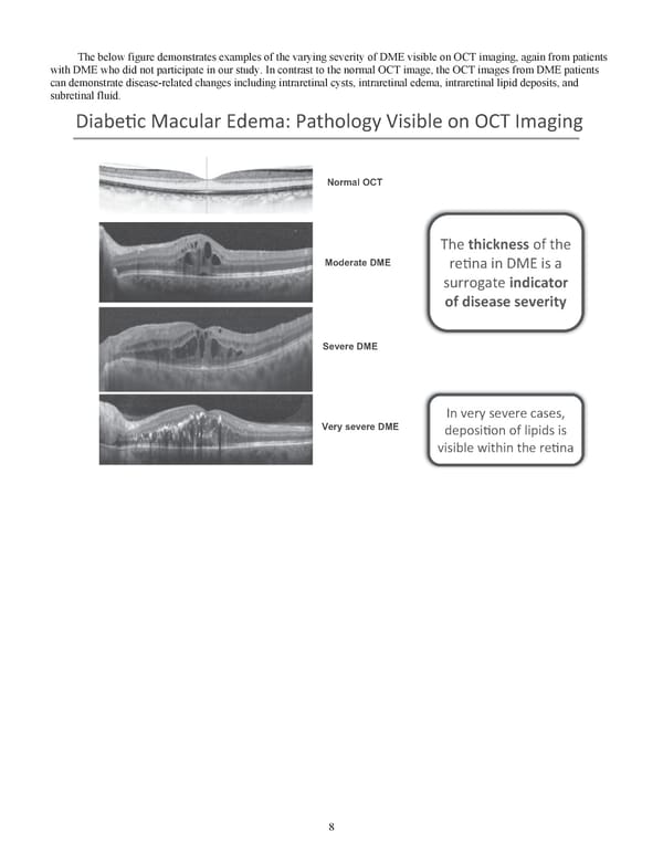
8 The below figure demonstrates examples of the varying severity of DME visible on OCT imaging, again from patients with DME who did not participate in our study. In contrast to the normal OCT image, the OCT images from DME patients can demonstrate disease-related changes including intraretinal cysts, intraretinal edema, intraretinal lipid deposits, and subretinal fluid. Diabe�c Macular Edema: Pathology Visible on OCT Imaging Normal OCT The thickness of the re�na in DME is a surrogate indicator of disease severity Moderate DME Severe DME Very severe DME In very severe cases, deposi�on of lipids is visible within the re�na
 Kodiak 2018 Annual Report Page 19 Page 21