
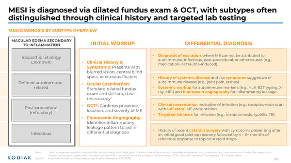
MESI is diagnosed via dilated fundus exam & OCT, with subtypes often distinguished through clinical history and targeted lab testing Note: 1 Used to evaluate anterior chamber cells, vitreous haze, and other signs of intraocular inflammation; 2 Identifies intraretinal cysts, subretinal fluid, and central sub foveal thickness; HLA = Human Leukocyte Antigen, ME = Macular Edema; MESI = Macular Edema Secondary to Inflammation, OCT = Optical Coherence Tomography, TB = Tuberculosis Source: American Academy of Ophthalmology; Expert Interviews (JUN 2025) • Clinical presentation indicative of infection (e.g., toxoplasmosis scar) with unilateral ME presentation • Targeted lab tests for infection (e.g., toxoplasmosis, syphilis, TB) • History of recent cataract surgery with symptoms presenting after an initial good post-op recovery followed by 4 – 6+ months of refractory response to topical steroid drops • History of systemic disease and / or symptoms suggestive of autoimmune disease (e.g., joint pain, rashes) • Systemic workup for autoimmune markers (e.g., HLA-B27 typing, X- ray, MRI) and fluorescein angiography for inflammatory leakage • Diagnosis of exclusion , where ME cannot be attributed to autoimmune, infectious, post-procedural, or other causes (e.g., medication- or trauma-induced) MACULAR EDEMA SECONDARY TO INFLAMMATION Idiopathic (etiology unknown) Defined autoimmune- related Post-procedural (refractory) Infectious INITIAL WORKUP DIFFERENTIAL DIAGNOSIS • Clinical History & Symptoms: Presents with blurred vision, central blind spots, or vitreous floaters • Ocular Examination: Standard dilated fundus exam and slit-lamp bio- microscopy 1 • OCT 2 : Confirms presence, location, and severity of ME • Fluorescein Angiography: Identifies inflammatory leakage pattern to aid in differential diagnosis MESI DIAGNOSIS BY SUBTYPE OVERVIEW 117
 Investor R&D Day Presentation Page 115 Page 117