
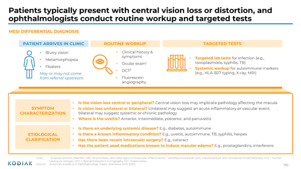
Patients typically present with central vision loss or distortion, and ophthalmologists conduct routine workup and targeted tests MESI DIFFERENTIAL DIAGNOSIS PATIENT ARRIVES IN CLINIC • Blurry vision • Metamorphopsia • Floaters May or may not come from referral upstream ROUTINE WORKUP • Clinical history & symptoms • Ocular exam 1 • OCT 2 • Fluorescein angiography TARGETED TESTS • Targeted lab tests for infection (e.g., toxoplasmosis, syphilis, TB) • Systemic workup for autoimmune markers (e.g., HLA-B27 typing, X-ray, MRI) SYMPTOM CHARACTERIZATION • Is the vision loss central or peripheral? Central vision loss may implicate pathology affecting the macula • Is vision loss unilateral or bilateral? Unilateral may suggest an acute inflammatory or vascular event; bilateral may suggest systemic or chronic pathology • Where is the uveitis? Anterior, intermediate, posterior, and panuveitis ETIOLOGICAL CLARIFICATION • Is there an underlying systemic disease? E.g., diabetes, autoimmune • Is there a known inflammatory condition? E.g., uveitis, autoimmune, TB, syphilis, herpes • Has there been recent intraocular surgery? E.g., cataract • Has the patient used medications known to induce macular edema? E.g., prostaglandins, interferons Note: 1 Evaluate anterior chamber cells, vitreous haze, and other signs of intraocular inflammation; 2 Identifies intraretinal cysts, subretinal fluid, and central sub-foveal thickness; HLA = Human Leukocyte Antigen, OCT = Optical Coherence Tomography, TB = Tuberculosis; Source: American Academy of Ophthalmology; Expert Interviews (JUN 2025) 116
 Investor R&D Day Presentation Page 114 Page 116