
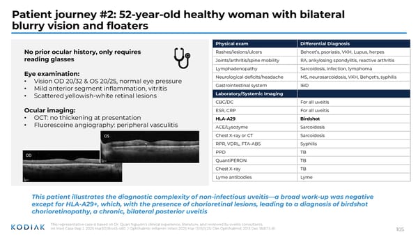
Patient journey #2: 52-year-old healthy woman with bilateral blurry vision and floaters No prior ocular history, only requires reading glasses Eye examination: • Vision OD 20/32 & OS 20/25, normal eye pressure • Mild anterior segment inflammation, vitritis • Scattered yellowish-white retinal lesions Ocular imaging: • OCT: no thickening at presentation • Fluoresceine angiography: peripheral vasculitis Physical exam Differential Diagnosis Rashes/lesions/ulcers Behcet’s, psoriasis, VKH, Lupus, herpes Joints/arthritis/spine mobility RA, ankylosing spondylitis, reactive arthritis Lymphadenopathy Sarcoidosis, infection, lymphoma Neurological deficits/headache MS, neurosarcoidosis, VKH, Behçet's, syphilis Gastrointestinal system IBD Laboratory/Systemic Imaging CBC/DC For all uveitis ESR, CRP For all uveitis HLA-A29 Birdshot ACE/Lysozyme Sarcoidosis Chest X-ray or CT Sarcoidosis RPR, VDRL, FTA-ABS Syphilis PPD TB QuantiFERON TB Chest X-ray TB Lyme antibodies Lyme This patient illustrates the diagnostic complexity of non-infectious uveitis—a broad work-up was negative except for HLA-A29+, which, with the presence of chorioretinal lesions, leading to a diagnosis of birdshot chorioretinopathy, a chronic, bilateral posterior uveitis This representative case is based on Dr. Quan Nguyen’s clinical experience, literature, and reviewed by uveitis consultants. Int Med Case Rep J. 2025 Mar30;18:445-460. J Ophthalmic Inflamm Infect 2025 Mar 13;15(1):25; Clin Ophthalmol. 2013 Dec 18;8:73-81 105
 Investor R&D Day Presentation Page 103 Page 105